The normal heart rhythm and anatomy
The normal heart rhythm and anatomy
To understand the different usages of cardiac devices, it is important to look at the anatomy of the heart and the normal heart rhythm first.
The heart is a muscular pump consisting of four chambers which circulate the blood through the whole body. The two upper chambers are called atria (right and left atrium) and receive the oxygenated blood from the lungs (left atrium) or deoxygenated blood from the rest of the body (right atrium). The two lower chambers are called ventricles (right and left ventricle) and pump the blood to the lungs (right ventricle) or to the rest of the body (left ventricle).
The contractions of the heart muscle are stimulated and synchronized by electrical signals which normally follow a specific electrical circuit within the heart, the conduction system. The electrical impulse begins in a group of heart cells at the top of the right atrium called sinus node, sometimes referred to as the body’s natural pacemaker. The electrical signals from the sinus node spread through both atria, making them pump. At the bottom of the right atrium is the atrioventricular node (AV node) which acts as a junction box. From here, the electrical signals are directed across the heart valves (which are like a layer of electrical insulation) through the His Bundle and into the bundle branches and His-Purkinje system. This network of fibers act like wires to rapidly spread the electrical impulse through both ventricles.
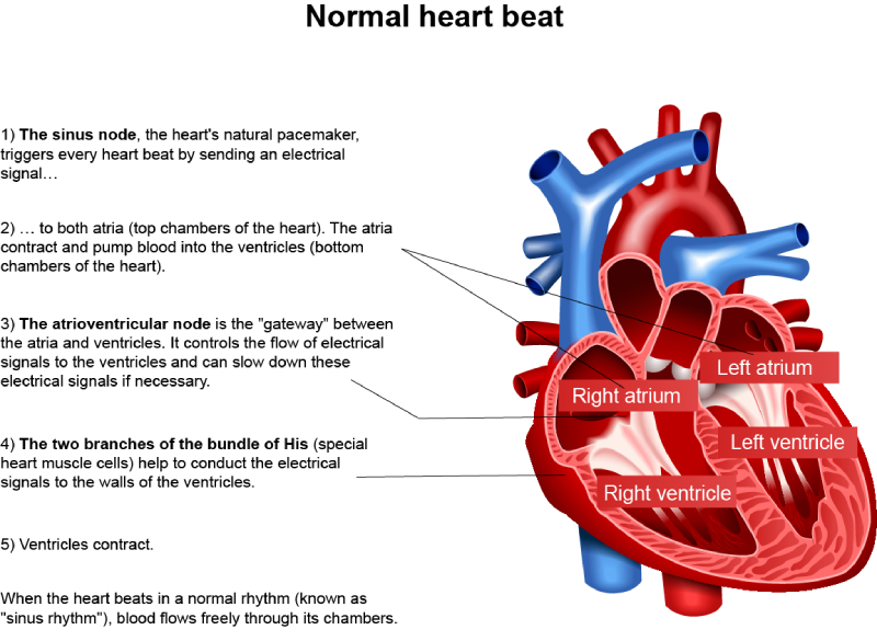
When the electrical signals stimulate the atria, both atria contract and pump the blood into the ventricles. When the electrical signals reach the ventricles, the right and left ventricles pump the blood into the lungs and rest of the body, respectively. This sequence is completed within one heartbeat.
FREQUENTLY ASKED QUESTIONS
What is a normal heart rhythm?
A normal heart rhythm, called sinus rhythm, is a steady heartbeat that originates in the sinoatrial (SA) node and ensures proper blood flow.
What is the role of the sinoatrial node?
The sinoatrial node, also called the heart’s natural pacemaker, initiates the electrical signals that regulate the heartbeat.
How does the heart’s electrical system work?
The heart’s electrical system generates signals that travel from the SA node to the atria, AV node, and ventricles to coordinate contractions.
What are the key parts of heart anatomy related to rhythm?
Key parts include the atria, ventricles, sinoatrial node, atrioventricular node, and the conduction pathways.
What does an ECG show about heart rhythm?
An electrocardiogram (ECG) shows the electrical activity of the heart and helps detect abnormal rhythms or arrhythmias.
What is the difference between a normal and abnormal heart rhythm?
A normal rhythm is steady and regular, while an abnormal rhythm (arrhythmia) can be irregular, too fast, or too slow.
What is normal heart anatomy?
The heart consists of four chambers (two atria and two ventricles) and is supported by valves and blood vessels for circulation.