The number of leads vary depending on the number of chambers of the heart that need to be treated. A pacemaker can have 1, 2 or 3 leads. Some modern pacemakers do not have leads in order to deliver the electrical signals to the heart muscles. These pacemakers are called leadless.
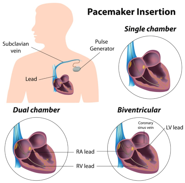
A single chamber pacemaker has only one electrode, which means that it delivers electrical impulses in only one chamber of the heart. The electrode can be placed in the right atrium of the heart (when the electrical signals begin in sinus node, but are slow) or in the right ventricle (when the heart rhythm is slow, but it does not begin in the sinus node, e.g. in cases of atrial fibrillation).
A dual chamber pacemaker has two leads which deliver electrical impulses to the right atrium and to the right ventricle of the heart. A dual chamber pacemaker is used when the electrical signals are blocked between the atrium and the ventricle (AV block) and it helps to coordinate the contraction of these two chambers.
A biventricular pacemaker, also called cardiac resynchronization therapy (CRT) has two or three electrodes, which deliver the electrical impulses to the right ventricle, left ventricle and right atrium. When the rhythm of the heart is atrial fibrillation, no electrode in the right atrium is required.
The role of a CRT device is to coordinate the contraction of the right and left ventricles, making each heartbeat more efficient.
Leadless pacemakers are small devices which consist of a pulse generator only, they have no leads. They are placed through the femoral vein at the top of the leg up into the right ventricle of the heart. They deliver electrical impulses through direct contact with the heart muscle. A leadless pacemaker can be used to stimulate only one chamber of the heart, the right ventricle. It is therefore restricted to those patients who only require single chamber pacing of the right ventricle.
A leadless pacemaker is a type of cardiac stimulator that does not require leads (wires) to regulate the cardiac rhythm. Unlike conventional pacemakers, whose generator is implanted in the pectoral region, under the skin, and connected to leads (electrical conductors) running through a vein down to the heart, leadless pacemakers are designed to operate without leads. Leadless pacemakers are compact (3 to 4 centimeters in length), self-contained devices implanted directly inside a heart chamber. They have a cylindrical metal design and are smaller than an AAA battery. With leadless pacemakers there is no need for leads since the pacemaker itself is in contact with the heart.
The leadless pacemaker is implanted into the right ventricle or the right atrium using a sheath (a long, thin tube) inserted via a vein in the groin (femoral vein). Once positioned, the device delivers electrical impulses to stimulate the heart and maintain a regular rhythm. The small device which is inside the right heart chambers contains the processor which operates pacing functions and the battery which gives the energy for operating the processor and pacing the heart. Being in contact with the inner surface of the heart chambers, it directly delivers electrical pulses with no need for leads. In case there are two devices, one in the atrium and the other in the ventricle, they communicate together to coordinate their actions. Therefore, a leadless pacemaker has a completely different design but functions exactly as a conventional pacemaker.
The implantation of a leadless pacemaker is a minimally invasive procedure performed in an electrophysiology laboratory. The procedure typically takes around 30-60 minutes to complete, though the duration may vary depending on the device type and individual anatomical factors. The process involves the following steps:
While leadless pacemakers offer certain advantages, they may not be suitable for all patients. Some factors to consider include:

Physiological pacing is defined as any form of cardiac pacing intended to restore or preserve synchrony of ventricular myocardium (cardiac muscle) contraction. This can be achieved in different ways, including conduction system pacing (CSP).

Figure 1. Pacemaker lead positioned in the conduction system.
In recent years, we have witnessed advancements in pacemaker implantation techniques. The placement of the ventricular electrode/lead in certain positions of the right ventricle can be associated with a condition known as pacemaker-induced cardiomyopathy, especially if the subject relies more on paced beats than on the spontaneous electrical activity of the heart. Abnormal heart contractions caused by the pacemaker, can lead to a worsening of cardiac function, potentially resulting in heart failure.
To counteract this, your physician may decide to implant the ventricular electrode/lead in a specific area of the electrical conduction tissue: the bundle of his or the left bundle branch, aiming to activate the heart’s electrical conduction tissue.

Figure 2. Pacemaker leads positioned in the bundle of his area (yellow circle) and in the left bundle branch area (yellow line).

Figure 3. Cardiac Magnetic Resonance Imaging showing the tip of the pacemaker lead in the interventricular septum close to the left ventricular endocardium (red arrow).
The procedure does not differ much from classic implantation except for additional stimulation to check the stimulation site and confirm that the right ventricular lead has been placed in the correct location and will stimulate the conduction system properly. Therefore, it will be normal during the implantation, if not performed under sedation, to hear the operators talking about values and measurements as they select the optimal area to implant the electrode to achieve the best electrical endpoint. This procedure may sometimes take longer than usual. Complications of the procedure may be slightly higher than for a conventional pacemaker, whereas the advantages seem to be valuable for the patients.

Figure 5. 12-lead electrocardiogram (ECG) with left bundle area pacing criteria

Figure 6. Transthoracic echocardiogram showing the tip of the pacemaker lead in the interventricular septum close to the left ventricular endocardium (red arrow).
A

B

Figure 7. X-RAY highlighting left ventricular lead positioned in the left bundle branch area (A) and in the conventional right ventricular apical area (B).
Pacemakers are classified as single-chamber, dual-chamber, and biventricular pacemakers. Single-chamber pacemakers stimulate one heart chamber, dual-chamber pacemakers stimulate both the atrium and ventricle, and biventricular pacemakers synchronize the ventricles for better heart function.
A biventricular pacemaker is designed for patients with heart failure. It coordinates contractions in both ventricles, improving the heart’s efficiency and symptoms of heart failure.
A pacemaker helps regulate a slow or irregular heartbeat, while an implantable defibrillator (ICD) monitors for dangerous arrhythmias and delivers shocks to restore normal rhythm.
Yes, pacemakers are typically implanted permanently, but they require battery replacements every 5-15 years depending on usage and device type.
A dual-chamber pacemaker sends electrical impulses to both the atrium and ventricle, ensuring they beat in coordination for optimal heart function.
A 3-lead pacemaker, also called a biventricular pacemaker, has leads that stimulate the right atrium, right ventricle, and left ventricle to improve heart synchronization in patients with heart failure.